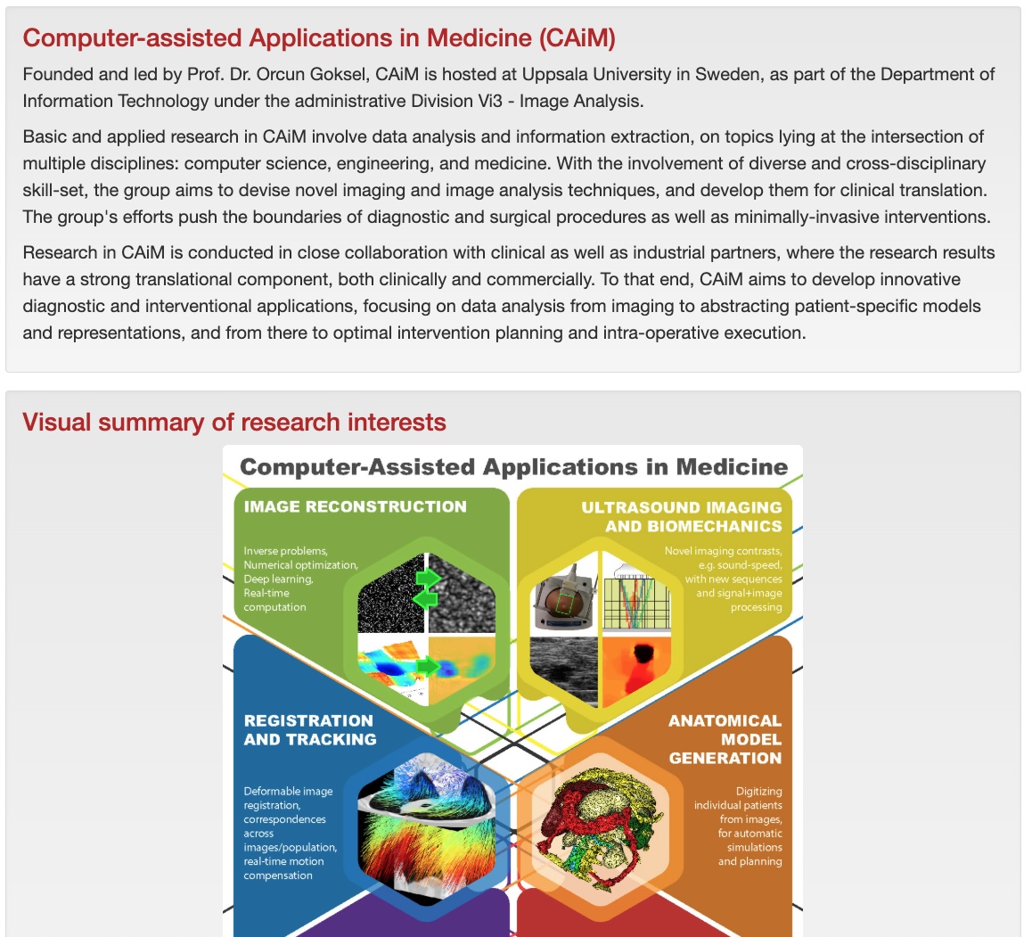
1
More information from the same exam
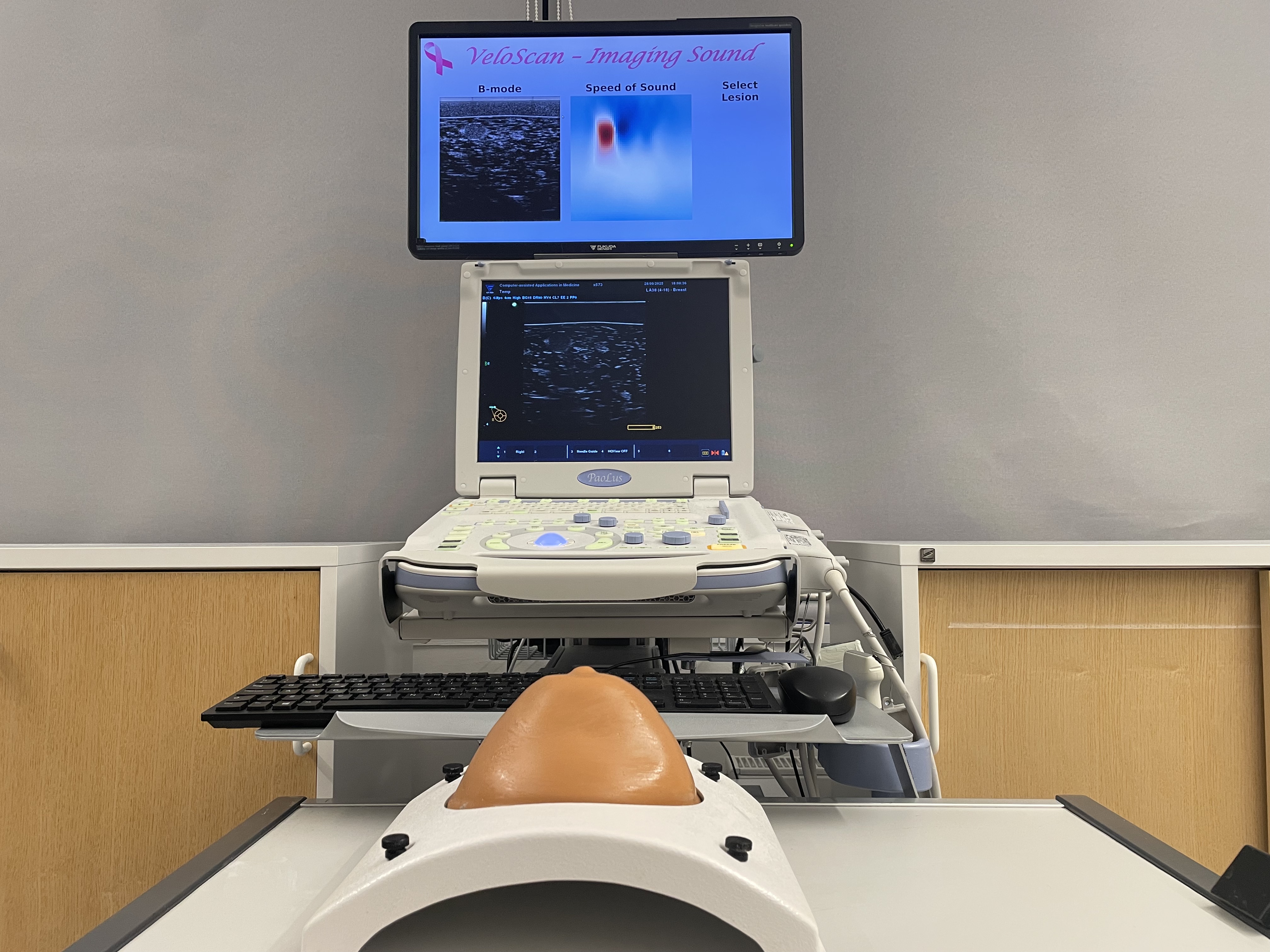
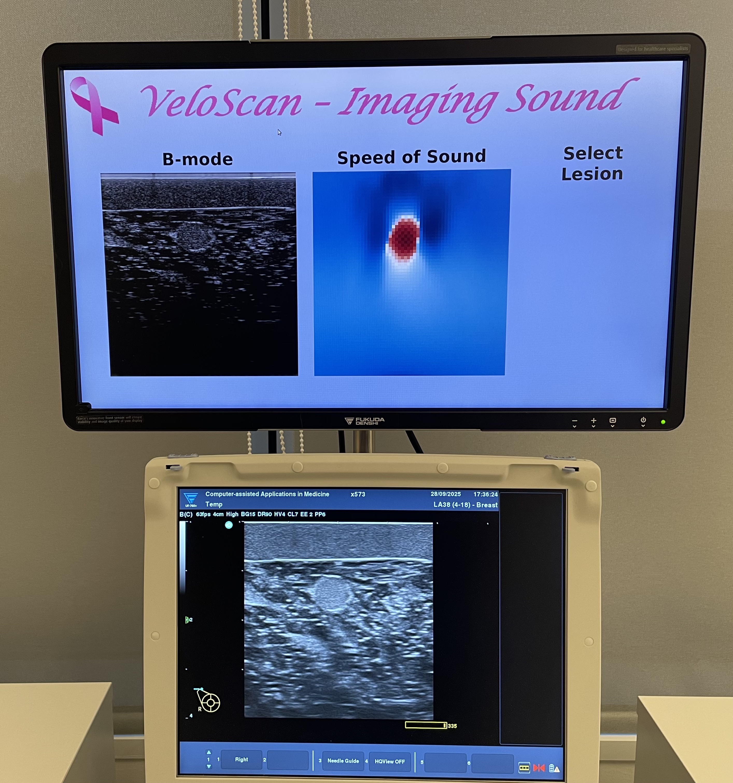
Sound-speed imaging adds tissue-property information to ultrasound, without changing the basic patient workflow.
Ultrasound is already used across healthcare, but it does not always provide enough information for confident decisions. Our approach uses sound-speed imaging to extract added insight into tissue properties from the same ultrasonic waves.
Ultrasound is safe, fast, affordable, and widely used. Yet conventional ultrasound mainly shows how strongly tissue reflects sound. That is not always enough to distinguish tissues or determine whether a change is disease-related.
Sound-speed imaging adds tissue-property information to ultrasound, without changing the basic patient workflow.
The added information can support diagnosis, triage, and follow-up in diseases where conventional ultrasound alone can be uncertain.
The approach is being developed for existing clinical ultrasound systems, including portable and handheld settings.
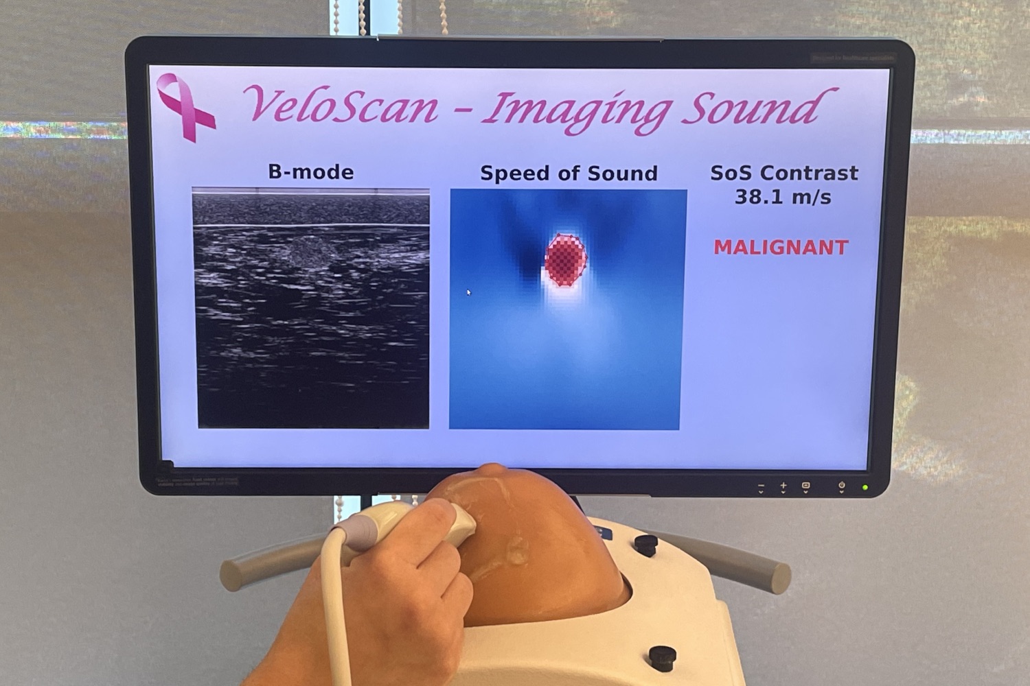
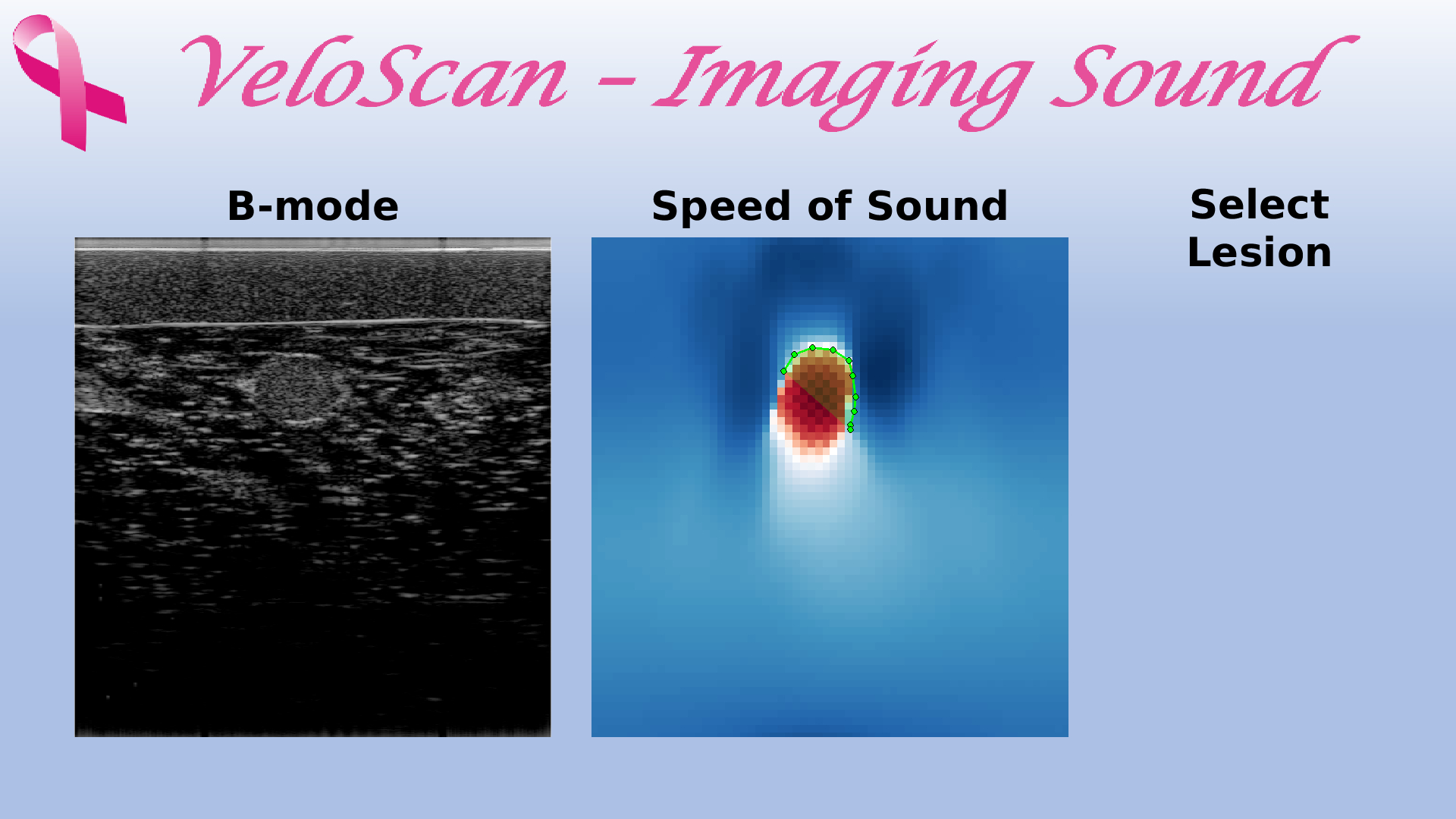
We image sound speed — how fast sound travels through tissue. Different tissues can transmit sound at different speeds. By turning this information into an image, ultrasound gains a new map of tissue content.
The aim is not to replace ultrasound, but to make it more informative: earlier diagnosis, better triage, fewer unnecessary examinations, and more equitable care.
The technology is being explored for clinical situations where accessible and objective tissue information could improve care pathways.
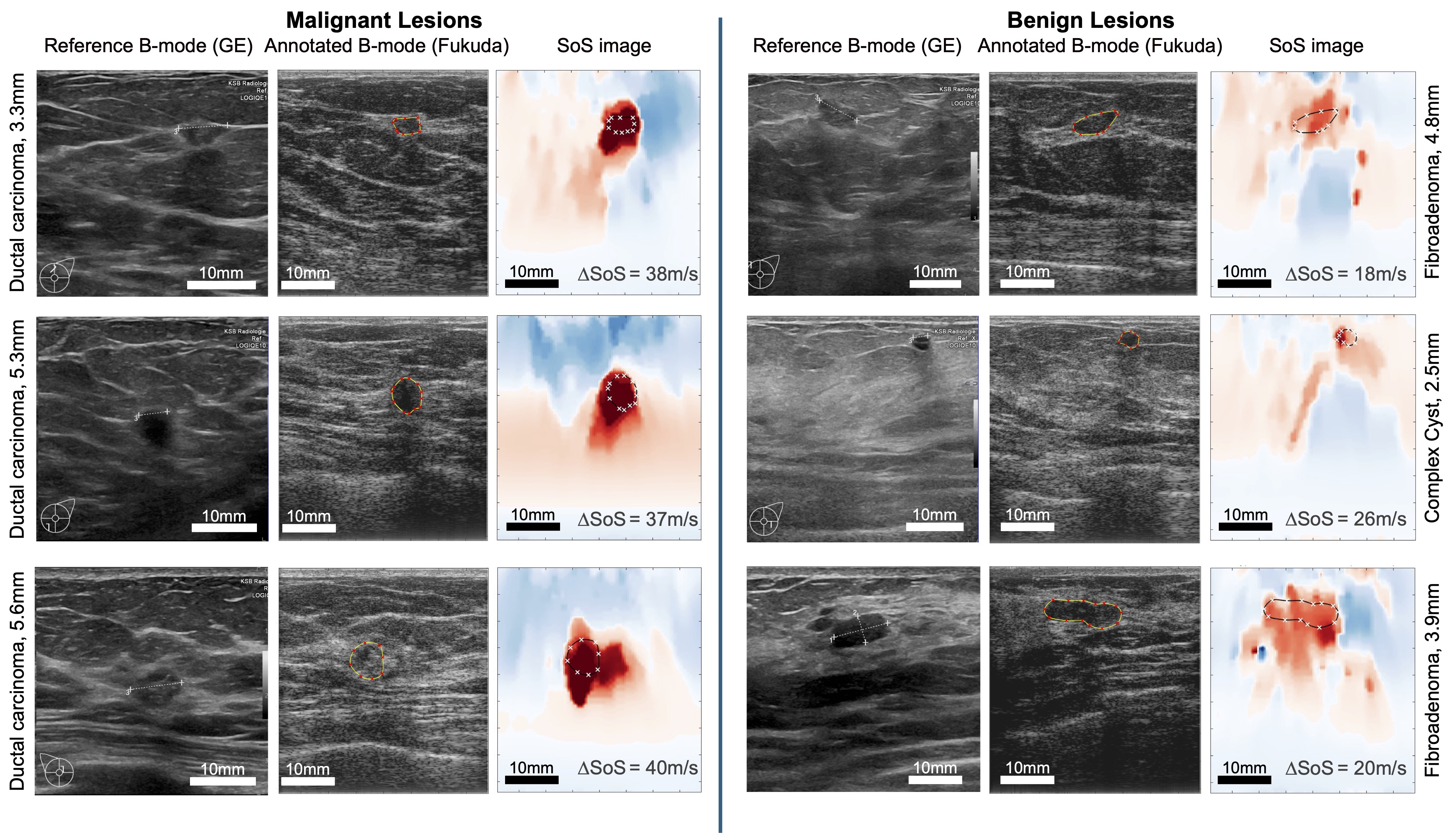
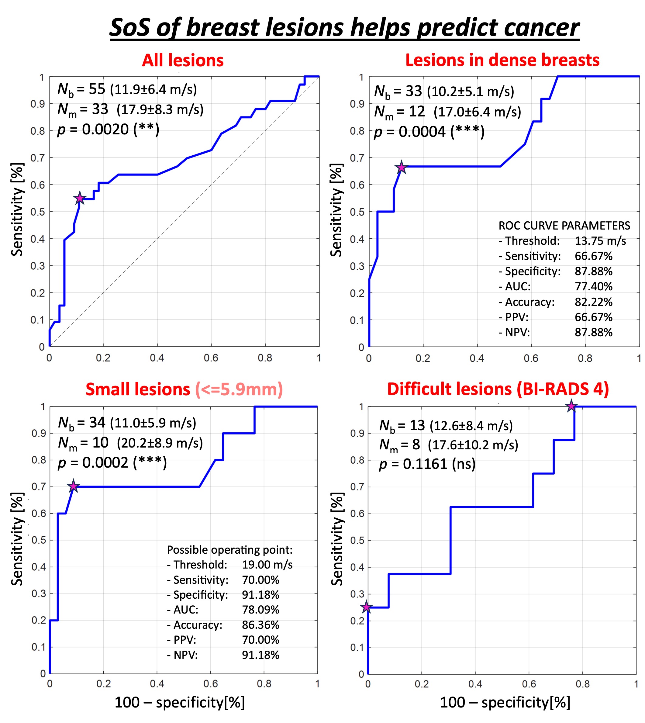
Clinical studies have shown promising results for breast cancer detection and breast density assessment, with the potential to support risk stratification and biopsy decisions.
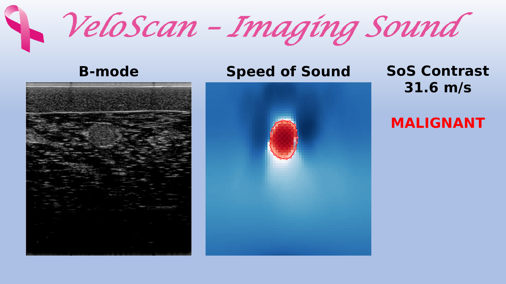
By adding information about tissue content, sound-speed imaging can support decisions where echo images alone are uncertain.
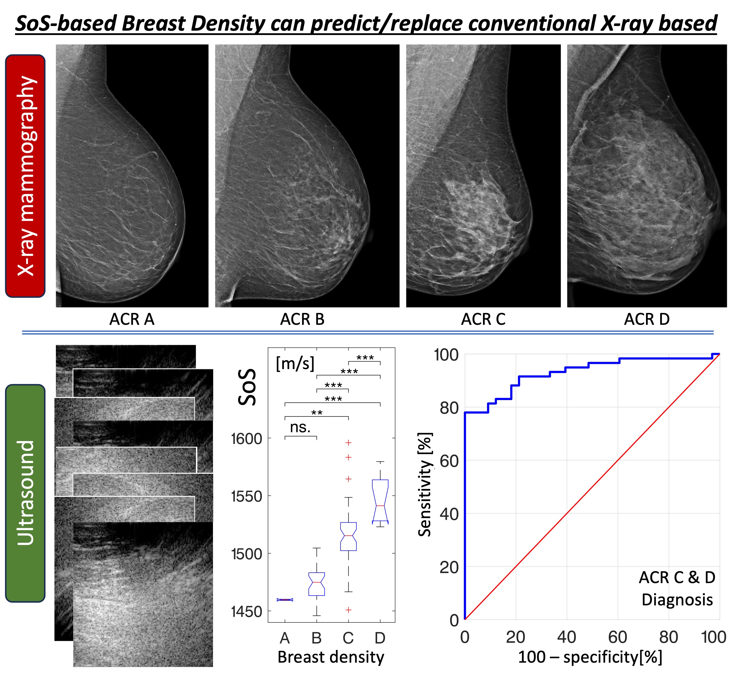
Quantitative tissue maps may support assessment of breast density and other factors relevant to risk stratification and monitoring.
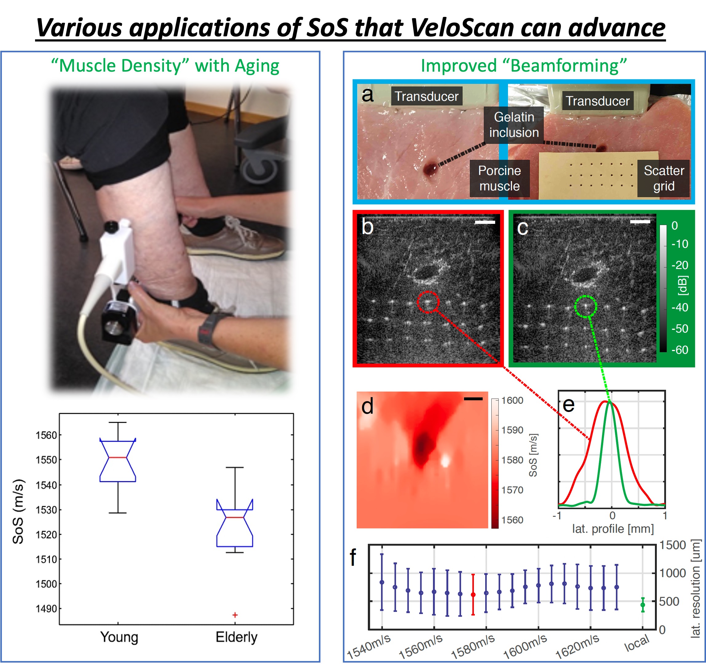
Future targets include fatty liver disease, muscular disease, and settings where accessible imaging can bring diagnostics closer to patients.
The research has progressed from method development to prototype and clinical evaluation. The next step is broader validation and integration into deployable systems.
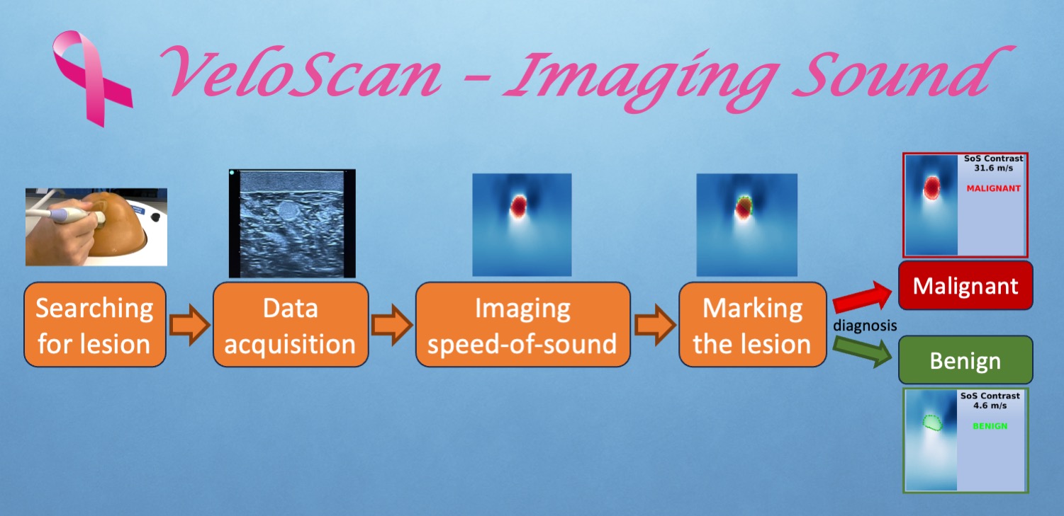
Convert raw ultrasound data into sound-speed images.
Evaluate diagnostic value with clinical partners.
Adapt the technology for existing ultrasound systems and workflows.
Work with healthcare and medtech partners toward everyday clinical use.
A compact gallery of project images and research figures. Select a tab to preview an image; click the large image to open it.
We seek healthcare providers, medtech companies, and innovation partners for clinical validation and product integration. The goal is to bring sound-speed imaging into practical use where it can improve diagnosis, triage, and access to care.
The work is led from the Computer-assisted Applications in Medicine group at the Department of Information Technology, Uppsala University, with clinical and research collaborators.


Selected links for background, recognition, and scientific evidence.
Accessible background on the project, the team, clinical prototype work, and how speed-of-sound imaging adds a new dimension to ultrasound.
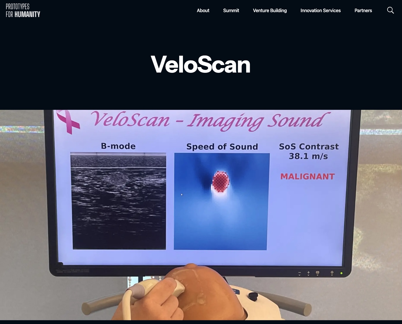
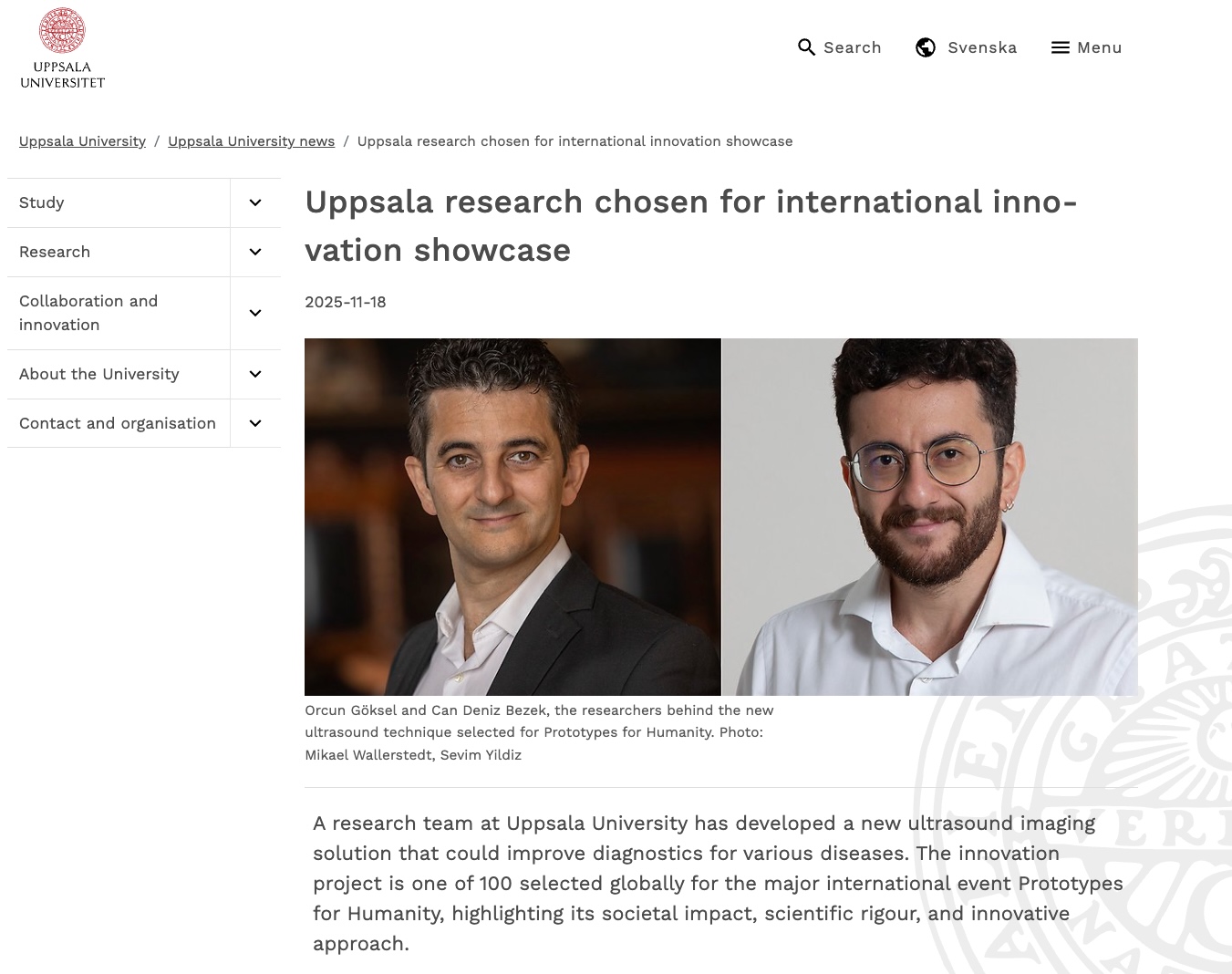
Project profile from Prototypes for Humanity, where the work was selected for the 2025 international innovation showcase.
Research-group context, publications, and contact information from Computer-assisted Applications in Medicine at Uppsala University.
Pulse-echo imaging of breast speed of sound as a potential biomarker for breast cancer